animal cell under microscope diagram
Animal cell under the microscope. We all do not forget that the human physique is quite problematic and a method I discovered to comprehend it is by.

Learn About The Plant Cell Science For Kids And Science Activities And Projects For Kids Cell Diagram Plant Cell Plant Cells Worksheet
We all keep in mind that the human physique is amazingly problematic and a method I learned to comprehend it is by means of the style of human anatomy diagrams.

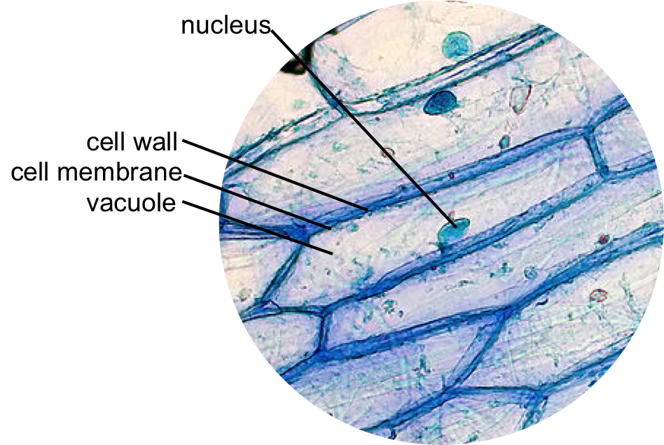
. To make observations and draw scale. Most of the cells are microscopic in size and can only be seen under the microscope. When observing onion cells there is the Cell Surface Membrane which is present in all living cells.
Cell Structure And Function. Observing a wide range of biological processes and animal cell under light microscope is easier due to advances in microscopic techniques. Students will observe cheek cells under a microscope.
Cell structure and organisation_notes igcsebiology dnl. We all keep in mind that the human body is quite intricate and a method I discovered to are aware of it is via the manner of. Below the basic structure is shown in the same animal cell on the left viewed with the light microscope.
While observing with tissues or on tissue. So lets begin by drawing a rough-oval shape. Its a thin slice.
Under the microscope animal cells appear different based on the type of the cell. Heres a diagram of a plant cell. It also shows the myoepithelial cells that surround each sweat gland of the animal skin.
One animal and one plant example given. Almost all animals and plants are made up of cells. Animal cells have a basic structure.
Tuesday April 20th 2021. A cell is the smallest functional and structural entity of life that it is easier observing animal cell under light microscope lensclutcolunch. But at the same time it is interpretive.
Most cells both animal and plant range in size between 1 and 100 micrometers and are thus visible only with the aid of a microscope. Get more skin-labeled diagrams on social media for anatomy learners. Record the microscope images using labelled diagrams or produce digital images.
Light and electron microscopes allow us to see inside cells. The shape of animal cells also varies with some being flat others oval or rod-shaped. Animal Cell Structure and Cell Organelles.
Skin cells under a microscope. The diagram is very clear and labeled. Living cells cannot be observed using an electron microscope because samples are placed in a vacuum.
There are various tasks done by a cell to complete them as the. Students will discover that their skin is made up of cells. Animal Cell Diagram Under Microscope Labeled.
We all keep in mind that the human physique is amazingly elaborate and one way I discovered to comprehend it is by way of the style of human anatomy diagrams. Students will discover that onions are made up of cells. Function cell does in the body.
As stated before animal cells are eukaryotic cells with a membrane-bound nucleus. Here is an electron micrograph of an animal cell with the labels superimposed. Generalized Structure of a Plant Cell Diagram.
A typical animal cell is 1020 μm in diameter which is about one-fifth the size of the smallest particle visible to the naked eye. Light is focused on the specimen by lenses in the condensor. When first examining cells or tissues with low power draw an image at.
However the internal structure and organelles are more or less similar. Thats the major difference between plant and animal cells under microscope. Animal cell project aviralgupta14.
The granulated area is the cell Cytoplasm while the huge round part is the Nucleus. Animal Cell Diagram Under Light Microscope. These are both specific typesof cells and from specific species.
This shows a generalized animal cell under a light microscope. As you can see in the above labeled plant cell diagram under light microscope there are 13 parts namely Cell membrane. Structure and Function Part 01.
A Dutch scientist named Anton van Leeuwenhoek observe these cells under the microscope and described bacterial cells and red blood cells of animals. A Diagram showing the light path in a compound microscope. Animal Cell Diagram Electron Microscope.
When observing onion cells there is the Cell Surface Membrane which is present in all living cells. We use microscope comprehensively in microbiology mineralogy cell biology biotechnology nano physics microelectronics pharmacology and forensics. Structure of animal cell and plant cell under microscope diagrams.
Anterior nasal structures medical images for. They are all typical elements of a cell. The first animal cell was observed under an optical microscope which clearly showed the nucleus and microfilament network in red and blue colors respectively.
Animal cell structure varies in different shapes. The cell was first discovered by an English natural philosopher Robert Hooke. Diagram Of Animal Cell Under Electron Microscope.
There are also more intriguing shapes such as curved spherical concave and rectangular. Students will observe onion cells under a microscope. Animal Cell Under Microscope.
Under the best conditions with violet light wavelength 0. Under the microscope animal cells appear different based on the type of the cell. After the invention of the microscope animal cells were first observed in the 17th century.
Function cell does in the body dictate the change and adaptation done by cell. An animal cell represents an eukaryotic cell in which true nucleus and other membrane-bound organelles such as mitochondria Golgi bodies and lysosomes are present. We all keep in mind that the human body is quite elaborate and a method I found out to comprehend it is by way of the manner of human.
Some may be oval or cylindrical shaped. A typical animal cell is 1020 μm in diameter which is about one-fifth the size of the smallest particle visible to the naked eye. Within the epidermis of a skin you will find squamous diamond-shaped and polyhedral cells under the light microscope.
Animal Cell Diagram Under Electron Microscope. Animal Cell Diagram Under Microscope.

Animal Cell Plant Cell Diagram Cell Diagram Animal Cell

Cellular Biology And Microscopy Ppt Download Animal Cell Structure Cell Organelles Electron Microscope

Related Image Electron Microscope Scientific Illustration Microscopy

Telophase Animal Cell Human Anatomy And Physiology Biology

Animal Cell Animal Cell Eukaryotic Cell Cell Biology

This Schematic Diagram Shows A Generic Animal Cell And The Organelles Including The Nucleus En Human Cell Diagram Human Cell Structure Animal Cells Worksheet

Image Result For Diagram Of Plant And Animal Cell Under Electron Microscope Celula Animal Ciencias Verdades Absolutas

Animal And Plant Cells Worksheet Inspirational 1000 Images About Plant Animal Cells On Pinterest Cells Worksheet Plant Cells Worksheet Animal Cell

The Great Story Iii An Animal Cell A Eukaryote Before The Electron Microscope The Complexity O Microscopic Photography Electron Microscope Science And Nature

Animal Cell Structure And Organelles With Their Functions Jotscroll Organelles Animal Cell Cell Diagram

Printable Labeled And Unlabeled Animal Cell Diagrams With List Of Parts And Definitions Animal Cell Cell Diagram Animal Cells Model

Organelle Structure And Function Plant Cell Animal Cell Plant And Animal Cells

Muppets Animal Drawing At Paintingvalley Com Explore Collection Of Muppets Animal Drawing Cell Diagram Animal Cell Structure Animal Cell

Animal Cell Model Diagram Project Parts Structure Labeled Coloring Animal Cell Plant And Animal Cells Animal Cells Model

Cells Under Electron Microscope Google Search Animal Cell Structure Cell Diagram Animal Cell

Year 11 Bio Key Points Cell Organelles And Their Function Cell Organelles Animal Cell Organelles

Draw It Neat How To Draw Animal Cell Animal Cell Drawing Animal Cell Human Cell Diagram

Epidermal Onion Cells Under A Microscope Plant Cells Appear Polygonal From The Cell Diagram Plant Cell Diagram Plant Cell